The Invisible Emergency You Can't Ignore
Retinal Detachment is a serious ocular emergency where the retina separates from its underlying supportive tissue and blood supply, potentially leading to permanent vision loss if not treated promptly. The thin layer of light-sensitive cells at the back of the eye pulls away from its normal position, disrupting the transmission of visual signals to the brain.
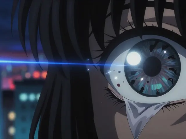
Imagine walking through your day and suddenly noticing a dark curtain sliding across your view. It isn't something you can rub away or blink off. If you ignore it, that shadow widens. Every hour you wait reduces your chance of saving good vision by approximately 5%. By the time many people finally call for help, the macula-the central part of the retina responsible for sharp vision-has already separated. This condition isn't a rare anomaly; it affects about 1 in 10,000 people annually in the general population. That number jumps significantly for anyone over 40.
We often talk about heart attacks or strokes being emergencies. But a retinal tear doesn't hurt much. There is no pain. Because there is no pain, patients often wait days. That delay is why so many end up with reduced acuity like 20/100 instead of the 20/25 vision they could have had if they acted fast. Knowing exactly what to look for is the difference between keeping your sight and facing blindness.
Six Warning Signs You Need to Spot Immediately
Your body gives you alerts before total separation happens. While a standard eye exam might catch early signs, most people discover issues when symptoms appear suddenly. According to clinical records from the American Academy of Ophthalmology, six specific signs consistently precede a full detachment.
- A sudden increase in floaters. You might see small dark spots or squiggly lines. If you report "a lot of new floaters" appearing within hours, take it seriously.
- Flashes of light (photopsias). These appear in peripheral vision, especially in dim light. Patients describe them like camera flashes without a bulb firing.
- A dark curtain. A shadow creeping across the visual field. NYU Langone Health ophthalmologists describe this as "the most urgent sign" requiring immediate attention.
- Sudden blurry vision. This occurs in 68% of documented cases. It feels like the lens of a camera has been smeared with grease.
- Loss of peripheral vision. In 73% of cases, side vision goes missing first. You might trip over objects that were previously visible in your corner eye.
- Changes in color perception. Particularly when the macula is affected, colors may lose their vibrancy instantly.
If you experience multiple symptoms at once, do not wait for morning appointments. Time is the one resource you cannot get back. A 2022 study in the Journal of VitreoRetinal Diseases showed 90% anatomical success rates when surgery occurs within 24 hours of symptom onset. Wait past that window, and the odds shift dramatically against you.
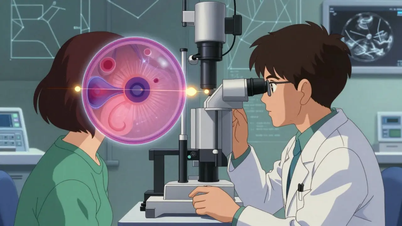
How Specialists Diagnose the Problem
General optometrists are great for glasses and routine checks, but diagnosing a detachment requires specialized gear. When you arrive at an emergency clinic, doctors perform a dilated fundus examination, which remains the gold standard. They put drops in your eyes to widen the pupil and shine a light into the back of your eye.
However, if the view is blocked by bleeding or cataracts, they can't see the retina directly. In those cases, they rely on B-scan Ultrasonography, a type of ultrasound specifically designed to image the posterior eye structures. This machine uses sound waves to build a picture of the back of the eye. Modern diagnostic setups usually include a slit lamp biomicroscope (like a Haag-Streit BQ 900) and an indirect ophthalmoscope with high-power lenses (such as a Keeler SuperQuad 20D). General ophthalmologists miss early detachments about 22% of the time compared to retinal specialists who miss only 5%.
You might also get Optical Coherence Tomography (OCT). This is a laser scan that creates cross-section images of the retina layers. It confirms whether fluid has slipped under the sensory retina. Proper diagnosis depends on having these tools available. If your local clinic lacks them, ask for a referral to a vitreoretinal specialist immediately.
Comparing Your Surgical Treatment Options
Once the diagnosis is confirmed, speed matters. Dr. Carl Regillo, Chief of Retina Service at Wills Eye Hospital, states that "every hour counts." There are three primary ways surgeons reattach the retina. Each has different success rates and side effects depending on your eye's anatomy.
| Treatment Method | Success Rate | Best For | Risks |
|---|---|---|---|
| Pneumatic Retinopexy | 70-80% | Single, superior breaks | High reoperation rate (30%) |
| Scleral Buckling | 85-90% | Young patients, lattice degeneration | Induced myopia (near-sightedness) |
| Vitrectomy | 90-95% | Complex cases, PVR | Cataract progression (70%) |
Pneumatic Retinopexy involves injecting a gas bubble into the eye. It works well for breaks located on the upper half of the retina. You stay face-down so gravity holds the gas bubble against the hole while it seals. The downside is it fails for lower breaks. About 30% of people need a second surgery later.
Scleral Buckling is an external procedure. The surgeon places a silicone band around the eye wall, indenting the sclera toward the retina. It preserves the natural lens longer than other methods. However, it changes the shape of the eye slightly, causing 1.5 to 2.0 diopters of induced myopia. You might need thicker glasses after recovery.
Vitrectomy is the most common approach, covering 65% of cases. Surgeons remove the gel inside the eye (vitreous) to relieve traction. They replace it with saline or gas. This gives the highest success rate for complex cases involving scar tissue (proliferative vitreoretinopathy). The catch is the risk of cataracts. In phakic patients (those with their natural lens), 70% develop cataracts within 2 years.
Newer technologies like the EVA Platform (DORC International) approved in January 2023 feature 27-gauge instruments that reduce surgical trauma. Smaller incisions mean faster healing times, making vitrectomy safer for older patients who might otherwise decline surgery due to fear of risks.
Recovery Realities: Positioning and Complications
Surviving the surgery is step one. Getting back to your life is step two. Post-operative care is grueling. If you had a gas bubble injected, you aren't done until the bubble absorbs. That usually takes 7 to 10 days. During this period, you must maintain a face-down position for roughly 50 hours a day. Some centers require 50 hours out of 24. Imagine sitting in a chair, chin resting on arms, staring at the floor.
This positioning causes significant neck strain. 41% of patients report discomfort during this phase. Some need home health assistance to eat or rest. It's critical to respect the positioning protocol. If the bubble isn't pressing against the retinal hole, the hole won't seal. Failure to comply leads to recurrence.
Other complications can arise months later. Elevated intraocular pressure happens in 25% of patients. Cataract progression is nearly guaranteed if you still have your natural lens. Recurrent detachment occurs in 5-15% of cases depending on the technique used. Long-term monitoring with your retinal specialist is essential.
Who Is Most at Risk?
You might wonder why this happened to you. Genetics play a role, but lifestyle and history matter too. High myopes (people with a prescription greater than -5.00D) have an annual incidence of 167 in 10,000. If you had cataract surgery, your risk rises to 0.5-2.0%. People with lattice degeneration have a 1% lifetime risk. Age is the biggest factor; incidence jumps to 20 in 10,000 among those over 40. The global market for treating this grew to $1.27 billion in 2022, driven mostly by aging populations needing these interventions.
Frequently Asked Questions
Is retinal detachment painful?
Usually, the condition itself is painless. Most people notice shadows, flashes, or floaters rather than physical pain. However, the surgery can cause some temporary discomfort or pressure sensations afterwards.
Can I fly after surgery?
If you have a gas bubble in your eye, you absolutely cannot fly until the bubble dissolves. Cabin pressure changes can cause the gas to expand, blinding you permanently. This can take several weeks. Saline-filled eyes do not have this restriction.
Will I regain perfect vision?
Outcomes depend on timing. If surgery happens before the macula detaches (macula-on), 90% of patients regain excellent vision. If the macula was detached (macula-off) before treatment, central vision loss is often permanent, though peripheral vision may return.
Does insurance cover retinal surgery?
Yes, Medicare and private insurers typically cover emergency repair. Average reimbursement for vitrectomy is around $7,200. Standard procedures are considered medically necessary treatments, not elective enhancements.
Can retinal detachment happen again?
Recurrence happens in 5-15% of cases. Factors like severe lattice degeneration or trauma increase the risk. Regular post-op exams monitor for tears in the remaining retina.